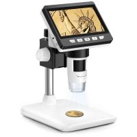
How Does Microscope Work?
Microscopes are essential tools in various fields of science and medicine, allowing us to observe objects and details that are otherwise invisible to the naked eye. Understanding how microscopes work can provide valuable insights into their applications and the principles behind their operation. This article will delve into the fundamental workings of microscopes, the different types available, and their practical uses.
The Basics of Microscopy

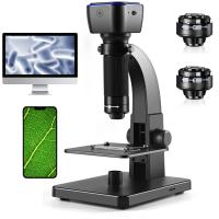
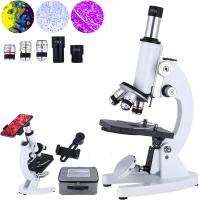
At its core, a microscope is an instrument designed to magnify small objects. The primary components of a basic optical microscope include the eyepiece (ocular lens), objective lenses, stage, light source, and focusing mechanisms. The process of magnification involves several steps:
1. Illumination: The light source, typically located beneath the stage, illuminates the specimen. In some microscopes, natural light or an external light source can be used.
2. Specimen Placement: The specimen is placed on a glass slide and positioned on the stage. The stage often has clips to hold the slide in place.
3. Objective Lenses: The objective lenses, located on a rotating nosepiece, are the primary magnifying lenses. They come in various magnifications, typically ranging from 4x to 100x.
4. Eyepiece: The eyepiece further magnifies the image produced by the objective lens. The total magnification is the product of the magnifications of the objective lens and the eyepiece.
5. Focusing: Coarse and fine focus knobs adjust the distance between the objective lens and the specimen, bringing the image into sharp focus.
Types of Microscopes
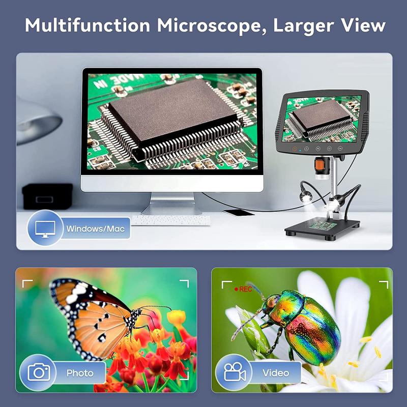
There are several types of microscopes, each designed for specific applications and offering different levels of magnification and resolution.
1. Optical Microscopes

Optical microscopes, also known as light microscopes, use visible light to illuminate the specimen. They are the most common type of microscope and include:
- Compound Microscopes: These microscopes use multiple lenses to achieve high magnification. They are ideal for viewing thin sections of specimens, such as cells and tissues.
- Stereo Microscopes: Also known as dissecting microscopes, they provide a three-dimensional view of the specimen. They are used for examining larger, opaque objects at lower magnifications.
2. Electron Microscopes

Electron microscopes use beams of electrons instead of light to achieve much higher magnifications and resolutions. There are two main types:
- Transmission Electron Microscopes (TEM): TEMs transmit electrons through a thin specimen, producing detailed internal images. They are used to study the ultrastructure of cells and viruses.
- Scanning Electron Microscopes (SEM): SEMs scan the surface of a specimen with a focused beam of electrons, creating detailed three-dimensional images. They are used to examine surface structures and topography.
3. Fluorescence Microscopes
Fluorescence microscopes use high-intensity light to excite fluorescent molecules within the specimen. These molecules emit light at different wavelengths, creating a highly detailed image. Fluorescence microscopy is widely used in biological research to study specific proteins, cells, and tissues.
Practical Applications of Microscopes
Microscopes have a wide range of applications across various fields, including biology, medicine, materials science, and forensic science.
1. Biological Research
In biological research, microscopes are indispensable for studying the structure and function of cells, tissues, and organisms. They allow scientists to observe cellular processes, identify microorganisms, and understand the complexities of biological systems. Techniques such as fluorescence microscopy enable researchers to label and visualize specific proteins and organelles within cells.
2. Medical Diagnostics
Microscopes play a crucial role in medical diagnostics. Pathologists use them to examine tissue samples and identify abnormalities, such as cancerous cells. Blood samples are analyzed under microscopes to detect infections, blood disorders, and other medical conditions. Electron microscopes are used to study viruses and other pathogens at the molecular level, aiding in the development of treatments and vaccines.
3. Materials Science
In materials science, microscopes are used to analyze the structure and properties of materials. Scanning electron microscopes (SEMs) provide detailed images of material surfaces, revealing information about their composition, texture, and defects. Transmission electron microscopes (TEMs) are used to study the internal structure of materials at the atomic level, which is essential for developing new materials and improving existing ones.
4. Forensic Science
Forensic scientists use microscopes to examine evidence from crime scenes. Microscopic analysis can reveal details about fibers, hair, soil, and other trace evidence that can link suspects to crimes. Scanning electron microscopes (SEMs) are particularly useful for analyzing gunshot residue, tool marks, and other forensic evidence.
Advanced Microscopy Techniques
Advancements in microscopy have led to the development of specialized techniques that provide even greater insights into the microscopic world.
1. Confocal Microscopy
Confocal microscopy uses laser light to scan specimens and create high-resolution, three-dimensional images. It eliminates out-of-focus light, resulting in clearer images of thick specimens. This technique is widely used in cell biology and neuroscience.
2. Super-Resolution Microscopy
Super-resolution microscopy techniques, such as STED (Stimulated Emission Depletion) and PALM (Photoactivated Localization Microscopy), break the diffraction limit of light, allowing scientists to observe structures at the nanometer scale. These techniques have revolutionized our understanding of cellular processes and molecular interactions.
3. Cryo-Electron Microscopy
Cryo-electron microscopy (cryo-EM) involves freezing specimens at extremely low temperatures and imaging them with an electron microscope. This technique preserves the native structure of biological molecules and complexes, providing detailed insights into their function. Cryo-EM has become a powerful tool in structural biology.
Microscopes are indispensable tools that have transformed our understanding of the microscopic world. From the basic optical microscope to advanced electron and super-resolution microscopes, these instruments have opened up new frontiers in science and medicine. By magnifying and revealing the intricate details of cells, tissues, materials, and forensic evidence, microscopes continue to play a vital role in research, diagnostics, and technological advancements.
Understanding how microscopes work and their various applications can help us appreciate the profound impact they have on our lives. Whether you are a student, researcher, or simply curious about the microscopic world, the study of microscopy offers a fascinating glimpse into the unseen and the unknown.


